
It is estimated that NF1 affects approximately 1 in 3000 people worldwide.1,2
PN, benign nerve sheath tumors, are a common manifestation of NF1 that may progress over time.2


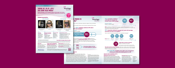
PN of any size and location can cause significant clinical complications that interrupt daily life7
‡Image reproduced from Gross AM, et al. 20184 (https://doi.org/10.1093/neuonc/noy067), public domain in the US.
§Image reproduced from Antônio JR, 20139 (http://dx.doi.org/10.1590/abd1806-4841.20132125), made available under the terms of the Creative Commons Attribution License (https://creativecommons.org/licenses/by/4.0/).


- Chronic, episodic, or both16
- Diffuse or localized to the PN site16
#Study of 60 children and adolescents with NF1 PN aged 6 to 18 years who were enrolled in a natural history protocol at the NCI. Caregivers and adolescents completed the IPI and BASC-II-P scales assessing the impact of NF1 PN.17
||CASSIOPEA was a retrospective study of 78 French children (aged ≥3 to <18 years) with NF1 PN referred to a specialist NF1 center or whose case underwent MDT review, confirming that the symptomatic PN was inoperable. The study was conducted between January 1, 2013, and December 31, 2019. A key exclusion criterion was prior or ongoing treatment with a MEK inhibitor as the study aimed to provide information on the natural history of NF1.18
- Proximity to vital organs
- Poorly defined margins
- Challenging locations such as the head, neck, spine, or trunk
- Hypervascularity, which can increase the risk of bleeding during surgery
- Delayed healing
- Bleeding
- Hematomas
- Necrosis
- Permanent neurological deficits
- Functional impairment
- Nerve damage
††In the 2012 study by Prada CE et al, subtotal resection was defined as 50% to 80% excision.6
Referral Brochure
Learn more about the impact of NF1 PN on your patients and treatment with Koselugo in our Referral Brochure.
Find Out More
Discover how Koselugo could help your patients with NF1 PN.
Adult Data
Pediatric Data

IMPORTANT SAFETY INFORMATION
WARNINGS AND PRECAUTIONS
Left Ventricular Dysfunction. Koselugo can cause cardiomyopathy, defined as a decrease in left ventricular ejection fraction (LVEF) ≥10% below baseline. In the pediatric safety pool, Grade 2 LVEF decrease occurred, as well as decreased LVEF of ≥20% resulting in dose interruption and dose reduction. The median time to first occurrence of LVEF decrease was approximately 12 months. In the adult population, Grade 2 LVEF decrease occurred, with decreased LVEF resulting in dose interruption. The median time to first occurrence of LVEF decrease was approximately 4 months. Assess ejection fraction by echocardiogram prior to initiating treatment, every 3 months during the first year of treatment, every 6 months thereafter, and as clinically indicated. Withhold, reduce dose, or permanently discontinue Koselugo based on severity of adverse reaction. In patients who interrupt Koselugo for decreased LVEF, obtain an echocardiogram or a cardiac MRI every 3 to 6 weeks until resolution. Upon resolution of decreased LVEF, obtain an echocardiogram or a cardiac MRI every 2 to 3 months.
Ocular Toxicity. Koselugo can cause ocular toxicity, including retinal vein occlusion (RVO), retinal pigment epithelial detachment (RPED), and blurred vision. In the pediatric safety pool, blurred vision, photophobia, cataracts, ocular hypertension, and retinal tear occurred. Blurred vision resulted in dose interruption. RPED occurred in the pediatric population during treatment with Koselugo and resulted in permanent discontinuation. In the adult population, blurred vision and vitreous floaters occurred in patients receiving Koselugo. Conduct ophthalmic assessments prior to initiating Koselugo, at regular intervals during treatment, and for new or worsening visual changes. Permanently discontinue Koselugo in patients with RVO. Withhold Koselugo in patients with RPED, conduct ophthalmic assessments every 3 weeks until resolution, and resume Koselugo at a reduced dose.
Gastrointestinal Toxicity. Koselugo can cause gastrointestinal toxicities, including diarrhea and colitis. In the pediatric safety pool (N=134), diarrhea occurred in 59% of patients, in addition to diarrhea resulting in permanent discontinuation and dose interruption. In the adult population (N=71), diarrhea occurred in 42% of patients who received Koselugo, in addition to diarrhea resulting in dose interruption. The median time to first onset of diarrhea was approximately 2 months in the pediatric safety pool and 1 month in the adult population. Advise patients to start an anti-diarrheal agent (eg, loperamide) and to increase fluid intake immediately after the first episode of diarrhea. Withhold, reduce dose, or permanently discontinue Koselugo based on severity of adverse reaction.
Skin Toxicity. Koselugo can cause severe rashes, including dermatitis acneiform. In the pediatric safety pool (N=134), rash occurred in 68% of patients. The most frequent rashes included dermatitis acneiform (47%) and maculopapular rash (31%). Pruritus, alopecia, and eczema occurred. In the adult population (N=71), rash occurred in 85% of patients who received Koselugo. The most frequent rash included dermatitis acneiform (66%). Alopecia and pruritus occurred in patients who received Koselugo. Grade 3 rash and rash resulting in dose interruption and dose reduction occurred in both the pediatric safety pool and the adult population. Permanent discontinuation also occurred in the adult population. Monitor for severe skin rashes. Withhold, reduce dose, or permanently discontinue Koselugo based on severity of adverse reaction.
Increased Creatine Phosphokinase (CPK). Koselugo can cause increased CPK, myalgia, and rhabdomyolysis. In the pediatric safety pool (N=134), increased CPK, based on laboratory data, occurred in 73% of patients, including Grade 3 or 4. In the adult population (N=71), increased CPK, based on laboratory data, occurred in 70% of patients who received Koselugo, including Grade 3 or 4. Increased CPK resulted in dose interruption and dose reduction in both the pediatric safety pool and adult population. Increased CPK concurrent with myalgia occurred in both populations, including one patient who permanently discontinued Koselugo for myalgia in the pediatric safety pool. Obtain serum CPK prior to initiating Koselugo, periodically during treatment, and as clinically indicated. If increased CPK occurs, evaluate for rhabdomyolysis or other causes. Withhold, reduce dose, or permanently discontinue Koselugo based on severity of adverse reaction.
Increased Levels of Vitamin E and Risk of Bleeding (Koselugo Capsules). Koselugo capsules contain vitamin E, which can inhibit platelet aggregation and antagonize vitamin K-dependent clotting factors. Supplemental vitamin E is not recommended if daily vitamin E intake (including the amount of vitamin E in Koselugo and supplement) will exceed the recommended or safe limits due to increased risk of bleeding. An increased risk of bleeding may occur in patients who are co-administered vitamin-K antagonists or anti-platelet antagonists with Koselugo capsules. Monitor for bleeding in these patients and increase international normalized ratio (INR) monitoring in patients taking a vitamin-K antagonist. Perform anticoagulant assessments more frequently and adjust the dose of vitamin-K antagonists or anti-platelet agents as appropriate. Koselugo oral granules do not contain vitamin E.
Embryo-Fetal Toxicity. Koselugo can cause fetal harm when administered during pregnancy. In animal studies, administration of selumetinib to mice during organogenesis caused reduced fetal weight, adverse structural defects, and effects on embryo-fetal survival at approximate exposures >5 times the human exposure at the clinical dose of 25 mg/m2 twice daily. Advise pregnant women of the potential risk to a fetus. Advise females of reproductive potential and males with female partners of reproductive potential to use effective contraception during treatment with Koselugo and for 1 week after the last dose.
ADVERSE REACTIONS
Common adverse reactions ≥40% in pediatric patients include vomiting, diarrhea, increased CPK, dry skin, paronychia, nausea, dermatitis acneiform, and pyrexia.
Common adverse reactions ≥40% in adult patients include rash (all), dermatitis acneiform, and diarrhea.
DRUG INTERACTIONS
Effect of Other Drugs on Koselugo
Concomitant use of Koselugo with a strong or moderate CYP3A4 inhibitor or fluconazole increased selumetinib plasma concentrations, which may increase the risk of adverse reactions. Avoid coadministration with Koselugo. If coadministration cannot be avoided, reduce Koselugo dosage.
Concomitant use of Koselugo with a strong or moderate CYP3A4 inducer decreased selumetinib plasma concentrations, which may reduce Koselugo efficacy. Avoid concomitant use with Koselugo.
SPECIAL POPULATIONS
Pregnancy & Lactation. Verify the pregnancy status of patients of reproductive potential prior to initiating Koselugo. Due to the potential for adverse reactions in a breastfed child, advise patients not to breastfeed during treatment with Koselugo and for 1 week after the last dose.
To report SUSPECTED ADVERSE REACTIONS, contact AstraZeneca at 1-800-236-9933 or at https://us-aereporting.astrazeneca.com or FDA at 1-800-FDA-1088 or www.fda.gov/medwatch.
INDICATIONKOSELUGO is indicated for the treatment of adult and pediatric patients 1 year of age and older with neurofibromatosis type 1 (NF1) who have symptomatic, inoperable plexiform neurofibromas (PN).
Please see full Prescribing Information for Koselugo (selumetinib) at https://alexion.com/Documents/koselugo_uspi.pdf.